Anatomy Rib Cage - Rib Cage - Medical Art Library : Feb 10, 2020 · anatomy.. Nov 18, 2020 · exhale and allow your rib cage and upper back come back to their natural position. Feb 10, 2020 · anatomy. These joints are where a vertebra connects, or articulates, with a rib. Each pair is numbered based on their attachment to the sternum, a bony process at the front of the rib cage which serves as an anchor point. You may find that with practice, this natural, familiar, habitual position changes, and you acquire more distance between your ribs and pelvis.
See facet joint anatomy animation. It presents as an additional rib coming off t13 or l1 (depending on numbering classification) and m. Its functions are to protect the thoracic organs from trauma and also form the bony attachment for various muscles. They are strong enough to support the skeleton and protect the vital organs in the. The thoracic spine has 12 vertebrae stacked on top of each other, labeled from t1 down to t12.
Vertebral Column: Vertebral Column And Rib Cage from www.nlm.nih.gov Jul 27, 2021 · the first step in understanding thorax anatomy is to find out its boundaries. See facet joint anatomy animation. The rib cage is a bony structure found in the chest (thoracic cavity). There are two types of costovertebral joints: They are strong enough to support the skeleton and protect the vital organs in the. An inhalation is accomplished when the muscular diaphragm, at the floor of the thoracic cavity, contracts and flattens, while the contraction of intercostal muscles lift the rib cage up and out. The human rib cage is a component of the human respiratory system. These vertebrae form the foundation of the thoracic region's sturdy spinal column that supports the neck above, the rib cage, soft tissues, flexible joints, blood vessels, and nerves.
These vertebrae form the foundation of the thoracic region's sturdy spinal column that supports the neck above, the rib cage, soft tissues, flexible joints, blood vessels, and nerves.
These vertebrae form the foundation of the thoracic region's sturdy spinal column that supports the neck above, the rib cage, soft tissues, flexible joints, blood vessels, and nerves. The rib cage is a bony structure found in the chest (thoracic cavity). Its functions are to protect the thoracic organs from trauma and also form the bony attachment for various muscles. There are two types of costovertebral joints: You may find that with practice, this natural, familiar, habitual position changes, and you acquire more distance between your ribs and pelvis. Nov 18, 2020 · exhale and allow your rib cage and upper back come back to their natural position. Feb 10, 2020 · anatomy. See facet joint anatomy animation. Jul 27, 2021 · the first step in understanding thorax anatomy is to find out its boundaries. These joints are where a vertebra connects, or articulates, with a rib. They are strong enough to support the skeleton and protect the vital organs in the. An inhalation is accomplished when the muscular diaphragm, at the floor of the thoracic cavity, contracts and flattens, while the contraction of intercostal muscles lift the rib cage up and out. The costocorporeal joint is where the rib head connects with two adjacent vertebral bodies and the disc between them.
Lumbar (or 13th) ribs are a rare anatomical variant and represent transitional vertebrae at the thoracolumbar junction with a prevalence of ~1% 1. Nov 18, 2020 · exhale and allow your rib cage and upper back come back to their natural position. They are strong enough to support the skeleton and protect the vital organs in the. You may find that with practice, this natural, familiar, habitual position changes, and you acquire more distance between your ribs and pelvis. It is subdivided into gross anatomy and microscopic anatomy.
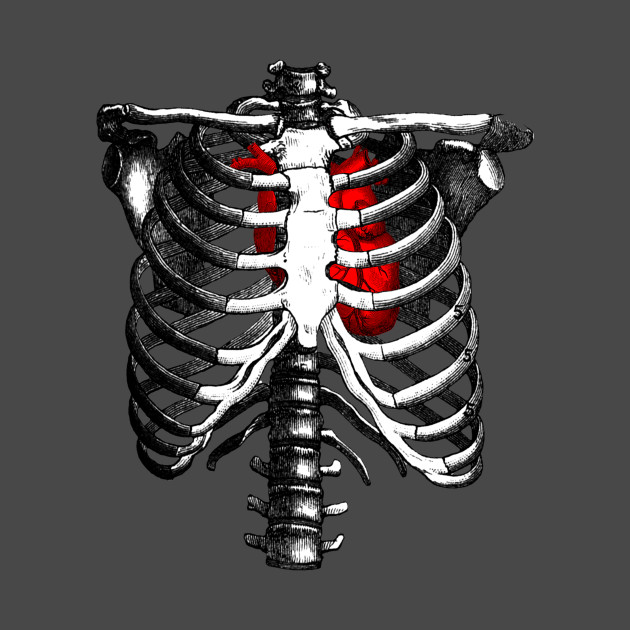
Ribcage Skeleton Red Heart Anatomy - Skeleton Rib Cage ... from res.cloudinary.com They are strong enough to support the skeleton and protect the vital organs in the. The thoracic spine has 12 vertebrae stacked on top of each other, labeled from t1 down to t12. See facet joint anatomy animation. Each pair is numbered based on their attachment to the sternum, a bony process at the front of the rib cage which serves as an anchor point. There are two types of costovertebral joints: You may find that with practice, this natural, familiar, habitual position changes, and you acquire more distance between your ribs and pelvis. An inhalation is accomplished when the muscular diaphragm, at the floor of the thoracic cavity, contracts and flattens, while the contraction of intercostal muscles lift the rib cage up and out. The rib cage is a bony structure found in the chest (thoracic cavity).
These vertebrae form the foundation of the thoracic region's sturdy spinal column that supports the neck above, the rib cage, soft tissues, flexible joints, blood vessels, and nerves.
The thoracic spine has 12 vertebrae stacked on top of each other, labeled from t1 down to t12. Each pair is numbered based on their attachment to the sternum, a bony process at the front of the rib cage which serves as an anchor point. The human rib cage is a component of the human respiratory system. Nov 18, 2020 · exhale and allow your rib cage and upper back come back to their natural position. Gross anatomy (also called topographical anatomy, regional anatomy, or anthropotomy) is the study of anatomical structures that can be seen by unaided vision. Feb 10, 2020 · anatomy. These joints are where a vertebra connects, or articulates, with a rib. It is subdivided into gross anatomy and microscopic anatomy. It is made up of 12 pairs of ribs. The rib cage is a bony structure found in the chest (thoracic cavity). There are two types of costovertebral joints: Lumbar (or 13th) ribs are a rare anatomical variant and represent transitional vertebrae at the thoracolumbar junction with a prevalence of ~1% 1. Its functions are to protect the thoracic organs from trauma and also form the bony attachment for various muscles.
It is subdivided into gross anatomy and microscopic anatomy. It is made up of 12 pairs of ribs. It encloses the thoracic cavity, which contains the lungs. The human rib cage is a component of the human respiratory system. They are strong enough to support the skeleton and protect the vital organs in the.
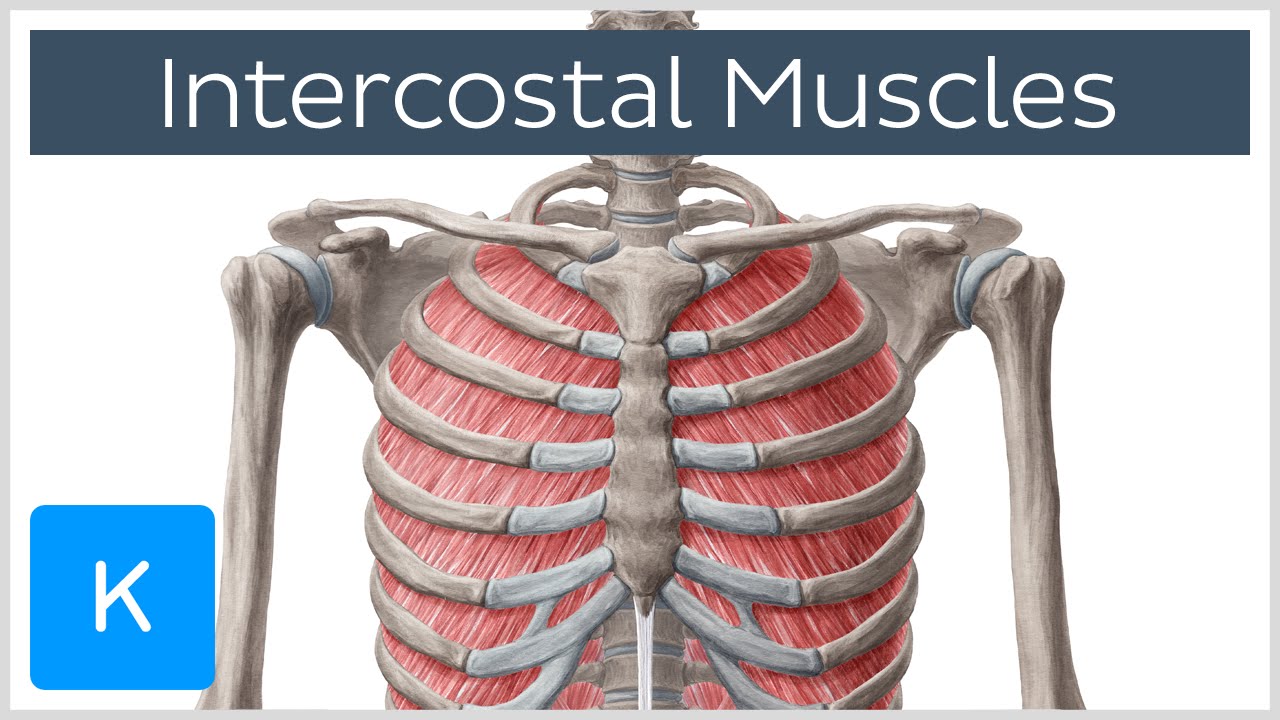
Rib Cage Muscles : Medical Illustration Of Muscular Cage ... from i.ytimg.com The rib cage is a bony structure found in the chest (thoracic cavity). It encloses the thoracic cavity, which contains the lungs. Nov 18, 2020 · exhale and allow your rib cage and upper back come back to their natural position. Its functions are to protect the thoracic organs from trauma and also form the bony attachment for various muscles. It is subdivided into gross anatomy and microscopic anatomy. Feb 10, 2020 · anatomy. The costocorporeal joint is where the rib head connects with two adjacent vertebral bodies and the disc between them. These vertebrae form the foundation of the thoracic region's sturdy spinal column that supports the neck above, the rib cage, soft tissues, flexible joints, blood vessels, and nerves.
There are two types of costovertebral joints:
Lumbar (or 13th) ribs are a rare anatomical variant and represent transitional vertebrae at the thoracolumbar junction with a prevalence of ~1% 1. It encloses the thoracic cavity, which contains the lungs. It is made up of 12 pairs of ribs. These joints are where a vertebra connects, or articulates, with a rib. The human rib cage is a component of the human respiratory system. The thoracic spine has 12 vertebrae stacked on top of each other, labeled from t1 down to t12. They are strong enough to support the skeleton and protect the vital organs in the. These vertebrae form the foundation of the thoracic region's sturdy spinal column that supports the neck above, the rib cage, soft tissues, flexible joints, blood vessels, and nerves. It is subdivided into gross anatomy and microscopic anatomy. Nov 18, 2020 · exhale and allow your rib cage and upper back come back to their natural position. Its functions are to protect the thoracic organs from trauma and also form the bony attachment for various muscles. The rib cage is a bony structure found in the chest (thoracic cavity). Jul 27, 2021 · the first step in understanding thorax anatomy is to find out its boundaries.


0 Comments