A Diagram Of Joints And Bones In The Human Body : Knee Pain Joint Bone Anatomy Joint Under The Microscope Technic Computer Wallpaper Human Png Pngwing / Describe the two ways joints are categorized in the human body.
A Diagram Of Joints And Bones In The Human Body : Knee Pain Joint Bone Anatomy Joint Under The Microscope Technic Computer Wallpaper Human Png Pngwing / Describe the two ways joints are categorized in the human body.. Identify and explain the 3 range of motions found in human joints. The long bones of the body contain many distinct regions due to the way in which they develop. Human skeleton, the internal skeleton that serves as a framework for the body. This diagram depicts bones of the human body.human anatomy diagrams show internal organs, cells, systems, conditions, symptoms and sickness information and/or tips for healthy living. Great for artists and students studying human anatomy.
The hip joint is one of the most important joints in the human body. There are two ways to categorize joints. The human skeletal system consists of all of the bones, cartilage, tendons, and ligaments in the body. Altogether, the skeleton makes up about 20 percent of a person's body weight. We'll go over the bones, joints, muscles, nerves, and blood vessels that make up the human arm.
How Many Muscles Are In The Human Body Plus A Diagram from i0.wp.com Human body muscles major muscles big muscles muscles of the. This diagram shows the six classes of movable joints in the human body. Identify and explain the 3 range of motions found in human joints. Aktuelle preise für produkte vergleichen! Side of skull (parietal bone) title: The epiphyseal plate of growing long bones and the first sternocostal joint that unites the first rib to the sternum are examples of synchondroses. It bears our body's weight and the force of the strong muscles of the hip and leg. This joint allows the head to rotate from left to right and back.
The long bones of the body contain many distinct regions due to the way in which they develop.
Über 7 millionen englischsprachige bücher. The skeleton of the human body is made out of bones and the cartilage supporting those bones. The bones of the leg are the femur, tibia, fibula and patella.the foot bones shown in this diagram are the talus, navicular, cuneiform, cuboid, metatarsals and calcaneus. There are five main shapes of bones: Joints in the human skeleton can be grouped by function (range of motion) and by. The patella and the pisiform bone of the carpals are the only sesamoid bones that are counted as part of the 206 bones of the body. When you finish with the above photo, head to the following model on posemanics and draw and label the linked model. Altogether, the skeleton makes up about 20 percent of a person's body weight. The epiphyseal plate of growing long bones and the first sternocostal joint that unites the first rib to the sternum are examples of synchondroses. Without your bones, you'd just be one big blob! Human skeleton, the internal skeleton that serves as a framework for the body. At a symphysis, the bones are joined by fibrocartilage. These muscles aid in the digestive.
The hip joint is one of the most important joints in the human body. Joints hold the skeleton together and support movement. Great for artists and students studying human anatomy. Long (such as the upper arm), short (such as the hand), flat (such as the ribs), irregular (such as the vertebrae) and sesamoid (such as the kneecap). The human skeletal system consists of all of the bones, cartilage, tendons, and ligaments in the body.
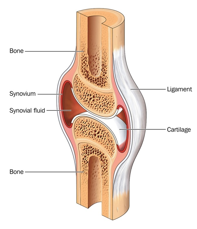
What Is Cartilage from www.news-medical.net The following activities will help your students understand and appreciate the work of their bones, muscles, and joints. Includes labeled human skeleton chart. Cartilage is an extremely flexible type of tissue, which is why it is located around joints. These muscles aid in the digestive. These muscles hold bones together, give the body its shape, and help move bones. This article is concerned primarily with the gross structure and the function of the skeleton of the normal. There are five main shapes of bones: The bones of the leg are the femur, tibia, fibula and patella.the foot bones shown in this diagram are the talus, navicular, cuneiform, cuboid, metatarsals and calcaneus.
Great for artists and students studying human anatomy.
These muscles aid in the digestive. #human body bones muscles and joints #human body series bones muscles and joints #human body with muscles and bones #the human body bones joints and muscles #the major bones and muscles in the human body This framework consists of many individual bones and cartilages.there also are bands of fibrous connective tissue—the ligaments and the tendons—in intimate relationship with the parts of the skeleton. We also have more than _____ muscles. It bears our body's weight and the force of the strong muscles of the hip and leg. Cartilage and bones are both connective tissues, and cartilage can be made out of different ratios of elastin or collagen. Ligaments are fibrous strands that connect bones. Human skeleton, the internal skeleton that serves as a framework for the body. The arm is one of the body's most complex and frequently used structures. Human body muscles major muscles big muscles muscles of the. There are two ways to categorize joints. A pivot joint allows one bone to rotate around another. Describe the two ways joints are categorized in the human body.
There are five main shapes of bones: Cartilage is an extremely flexible type of tissue, which is why it is located around joints. This diagram shows the six classes of movable joints in the human body. At a synchondrosis, the bones are united by hyaline cartilage. They hold up your body, and along with your muscles, keep you moving.
Joints And Skeletal Movement Biology Ii from cnx.org The arm is one of the body's most complex and frequently used structures. Long (such as the upper arm), short (such as the hand), flat (such as the ribs), irregular (such as the vertebrae) and sesamoid (such as the kneecap). Yet the hip joint is also one of our most flexible joints and allows a greater range of motion than all other joints in the body except for the shoulder. The human skeletal system consists of all of the bones, cartilage, tendons, and ligaments in the body. When you finish with the above photo, head to the following model on posemanics and draw and label the linked model. These muscles aid in the digestive. At a symphysis, the bones are joined by fibrocartilage. Human skeleton, the internal skeleton that serves as a framework for the body.
Other sesamoid bones can form in the joints of the hands and feet, but are not present in all people.
This article is concerned primarily with the gross structure and the function of the skeleton of the normal. This joint allows the head to rotate from left to right and back. The patella and the pisiform bone of the carpals are the only sesamoid bones that are counted as part of the 206 bones of the body. There are two ways to categorize joints. Ball and socket joints, like your hip and shoulder. The human skeletal system consists of all of the bones, cartilage, tendons, and ligaments in the body. Bones store calcium and release it into the bloodstream when other parts of the body need it. Includes labeled human skeleton chart. The arm is one of the body's most complex and frequently used structures. Other sesamoid bones can form in the joints of the hands and feet, but are not present in all people. The long bones of the body contain many distinct regions due to the way in which they develop. The epiphyseal plate of growing long bones and the first sternocostal joint that unites the first rib to the sternum are examples of synchondroses. Human body muscles major muscles big muscles muscles of the.


0 Comments